UNC Research Core Facilities are shared services that enhance and expand the collaborative capabilities of Carolina’s research enterprise.
More than 140 core facilities at UNC-Chapel Hill offer a range of resources to the research community, including cutting-edge technologies, high-end instrumentation, technical support, and education. Administered by various schools, units, and departments across campus, they serve a variety of disciplines, enabling researchers to investigate and innovate in ways that wouldn’t be possible within the scope of their individual labs or projects.
Each year Carolina’s core facilities:
- assist 3,000+ researchers at UNC-Chapel Hill and 500+ outside of the University;
- support 2,000+ sponsored programs and over $1 billion in sponsored research activity;
- contribute to 1,000+ peer-reviewed scientific publications;
- provide consulting and technical services to 160+ universities, companies, and non-profits; and
- generate $40 million in revenue.
Here’s a look at four cores and the services they provide.
Chapel Hill Analytical and Nanofabrication Laboratory (CHANL)
The CHANL core helps researchers tailor materials and micro-sized structures to advance their studies. The applications for this technology are varied and include solar energy, liquid fuels, biotechnology, pharmacology, and soft materials. That means that every day is different for Technical Director Bob Geil.
“It’s a multifaceted job that is very hands-on one day and has me in front of a computer the next,” says Geil, who also manages the core’s cleanroom — a controlled environment that allows for uncontaminated processes. “I’m developing processes and training students, but also getting my hands dirty and fixing things.”
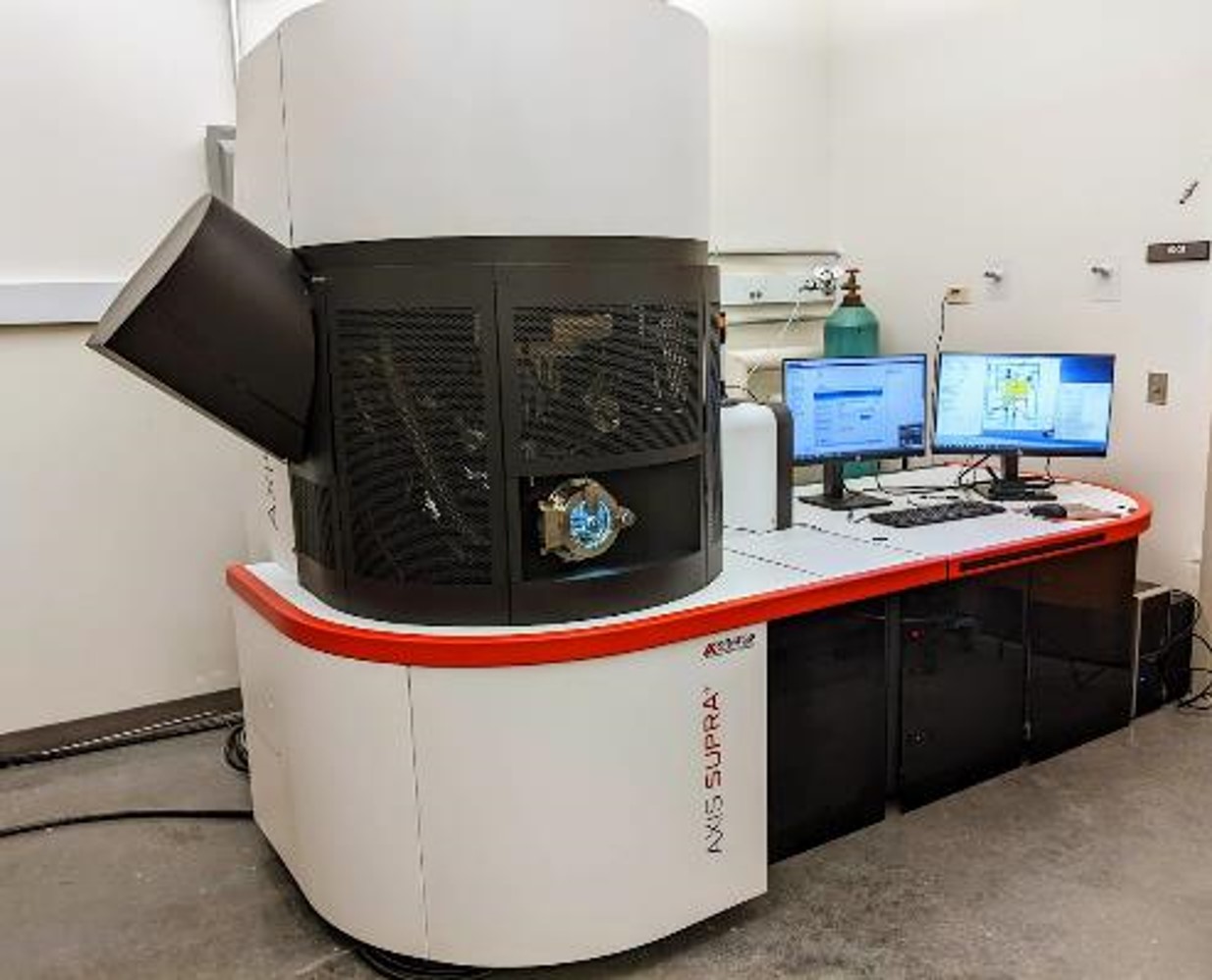
CHANL has more than 20 pieces of sophisticated research equipment that most individual labs could not afford on their own. They also have unique tools, like an X-ray photoelectron spectroscopy (XPS) system that can identify the elements of a material’s surface, such as chemical composition and oxidation state.
Other tools include a focused ion beam system, scanning and transmission electron microscope, and X-ray diffraction instrumentation. These, along with the XPS system, are used almost daily by researchers from Carolina and beyond.
“We have users from nearby universities and private industry,” Geil says. “We’re open to people with any skill level and provide training so users can operate the machines independently. And we don’t just support research, we provide education that can help young researchers advance their careers.”
CHANL is also part of the Research Triangle Nanotechnology Network (RTNN), which is funded by the NSF and is one of 16 U.S. sites included in the National Nanotechnology Coordinated Infrastructure. The RTNN is comprised of UNC-Chapel Hill, Duke University, and NC State, and together they promote research education, access, and innovation.
To work with CHANL, schedule a consultation to learn more about their services, cost, and training.
BeAM Design Innovation Hub
Born from Carolina’s BeAM Makerspaces, the BeAM Design Innovation Hub is led by Glenn Walters, who also serves as the senior technical director for the makerspace network. The hub provides engineering design and expertise for researchers who need a specific tool or part to conduct their studies but want help creating the final product.
“Even if you’re unsure of where to go with a request, it’s worth asking us because we can sometimes move quickly to fulfill a need,” Walters says. “And if we aren’t the right place for the request, we’ll connect you with the team on campus that can help.”
Walters also encourages the research community to take advantage of the makerspace network, which is free to use and provides training for all tools, including 3D printers, electronics, laser cutters, and metal and wood working machines.
“Once you have these tools under your belt, the way you think about solving problems changes,” Walters says. “Don’t worry about not knowing. It’s a welcoming space and our staff will teach you whatever you need to know.”
To set up a free consultation with the hub, contact Glenn Walters.
Clinical Genomic Analysis (GENYSIS)
As one of the newer core facilities at Carolina, GENYSIS aims to help researchers analyze whole exome and genome sequences — looking at a person’s entire genetic code. By working with existing core facilities, such as the BioSpecimen Processing Facility and High-Throughput Sequencing Facility, GENYSIS can provide a unique service for researchers looking to add clinical genetic sequencing, analysis, and results reporting to a study.
“We have combined capabilities that would be difficult to build in individual studies,” Director Tam Sneddon says. “We’re offering expertise in clinical genetics, laboratory genetics, genetic counseling, and bioinformatics, which are services that have been built up over a decade.”
The core works with researchers to comb through and interpret data, looking for genetic variants that could be useful for research and impactful to a person’s health.
“With this type of sequencing you get so much data,” says Kimberly Foss, assistant director of GENYSIS. “The variant interpretation is very complex, and so is clinical reporting. This group can help you with everything from writing the informed consent document to interpreting data and getting that information back to the participant and clinical team.”
Working with clinical teams guarantees that study participants understand their sequencing results and any health implications that could arise. The goal is more transparency and value for those who make the studies possible.
“A lot of times in the research world, something happens and it only stays in the research realm,” Foss says. “We make sure the patient gets a report and it goes in their medical record for more visibility over their long-term care. And we provide resources to the clinical team and the patient to inform care if more immediate action is needed.”
GENYSIS can also help researchers draft IRB language and facilitate patient consent, so contacting them early in the research process can be beneficial. The core can also aid ongoing projects with existing data. They offer free consultations.

Neuroscience Microscopy Core
The Neuroscience Microscopy Core within the School of Medicine is directed by cell biology and physiology assistant professor Michelle Itano. Itano specializes in the fluorescence microscope, a light microscope that uses various wavelengths — or colors — of light that interact with dyes or proteins.
Users “paint” specific parts of their specimens with dyes or proteins to illuminate specific structures. They can use multiple stains to label different parts of their specimen, creating an image similar to an abstract piece of art. This technique allows researchers to see events occurring in living animals, cells, and tissues.
These microscopes allow users to see with more detail than ever before. Previously, researchers needed access to multiple microscopes to capture a variety of images at different scales. Now, thanks to technological improvements, those scales can be achieved with the same instrument.
“You can go from a whole organism down to a single molecule and, with specialized techniques, get close to atomic levels of detail,” Itano says.
Itano teaches researchers how to take beautiful, complex images, assisting them with data analysis, image processing, problem-solving, and tailored advice for their specific projects — like how an IT professional might help you troubleshoot your computer problems.
She works with researchers all over the University, from neuroscientists to dentists to gastroenterologists to cancer researchers. She also collaborates with numerous organizations in N.C. and across the world — including the Light Microscopy Core Facility at Duke University, Harvard University’s MicRON core, BioImaging North America, and Global BioImaging.
To work with this core, submit a training request. This will initiate a consultation to discuss project goals, imaging needs, and the most appropriate systems for the study.
By Carleigh Gabryel, UNC Reseach